Electrospinning and Polymer Nanofibers: II. Properties and Applications
Personnel: L. Chen, J.L. Lowery, M. Ma, M. Wang, K.K. Gleason, R.M. Hill, D. Kaplan, S. Shortkroff
Sponsorship: NTC, US Army ISN (sponsor contact information)
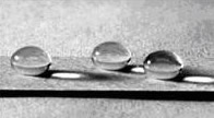
We are exploring the use of nanofibers for a variety of properties and applications. Topological features on the order of nanometers are known to be important to the superhydrophobic behavior of natureal and synthetic materials. We have developed two strategies to produce superhydrophobic textiles via electrospinning. The first strategy is to electrospin a block copolymer such as poly (styrene-b-dimethylsiloxane), in which the hydrophobic siloxane block spontaneously segregates to the fiber surface. Contact angle measurements indicated that the resultant nonwoven fibrous mats were superhydrophobic, with a contact angle of 163° (Figure 5) and contact angle hysteresis of 15°. The superhydrophobicity was attributed to the combined effects of surface enrichment in siloxane, as revealed by X-ray photoelectron spectroscopy, and surface roughness of the electrospun mat itself [Ma et al, Langmuir 2005 (in press)]. The second strategy is to combine electrospinning and initiated chemical vapor deposition (iCVD). A selected polymer was first electrospun into both beaded and bead-free fibers with different diameters and then coated with a thin layer of hydrophobic polymerized fluoroacrylate by iCVD. The hierarchical surface roughness inherent in the electrospun mats and the extremely low surface free energy of the coating layer obtained by iCVD yielded stable superhydrophobicity with a contact angle as high as 175° and a threshold sliding angle less than 2.5° for a 20 mg droplet. The coated mats were also shown to exhibit at least “Grade-8” oleophobicity (heptane-phobic). The systematic effect of fiber morphology on superhydrophobicity was investigated both experimentally and theoretically.
Submicron diameter polymer fibers provide a suitable mimic for the collagen fibrils that are ubiquitous in the extracellular matrix (ECM) of mammalian tissue. For applications involving fiber-based tissue engineering scaffolds, the nature of the pore structure has been found to increase with varying fiber size, making possible the creation of a three-dimensional mat with tunable material and structural characteristics. By implanting chondrocytes into the electrospun mats using fluid dialysis, chondrocyte cells survive and proliferate up to 4-weeks post-seeding in the electrospun scaffolds. By adding growth factors, these levels have been found to increase further. We seek to induce type II collagen production and eventually create cartilage tissue by allowing the electrospun polymer mats to degrade as natural ECM is produced by the implanted cells. Our all-aqueous process utilizing two-fluid electrospinning to produce solid, submicron diameter fibers of Bombyx mori silk could also have relevance for the manufacture of tissue engineering scaffolds.
To develop lightweight, strong and functional nanofibers, we have explored two approaches. The first is to integrate different nanoparticles such as nanoclay or magnetic nanoparticles into fiber systems to form nanocomposite fibers that exhibit improved thermal, mechanical or magnetic properties. In the area of nanocomposites, we have integrated two types of nanoparticles, nanoclay and magnetic nanoparticles, into the nanofiber matrix using electrospinning. We have investigated the relationships between processing, structure and properties of nanocomposite fibers. Clay nanocomposite fibers have shown improved thermal, mechanical properties over pure polymeric fibers, and magnetic nanofibers have shown “smart” strain-rate-dependent mechanical properties within an external magnetic field. The second approach is to process inherently strong natural and synthetic materials into fiber form. For this purpose, we developed an environmentally benign, entirely aqueous process utilizing two-fluid electrospinning to produce solid submicron diameter fibers of Bombyx mori silk. In as-spun nanofiber, silk fibroin preserves the random coil conformation due to the rapid fiber formation during electrospinning process. After methanol treatment, silk fibroin was converted from random coil into ß-sheet structure crystals. Evidence of the nanofibrils formed within as-spun nanofiber was observed and PEO phase are dispersed as small elongated islands within the silk fibroin matrix. The mechanical properties of single nanofiber were characterized by an AFM nanoindentation technique, and the results are consistent with the uniaxial tensile tests and morphological analysis.